
Presentation
Non specific knee pain and instability.
Patient Data
Age: 25 years
Gender: Female
From the case:
Cruciate ligaments of the knee
Show annotations
Download
Info
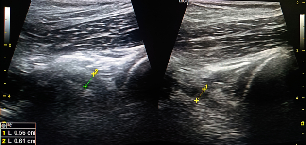
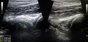
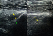
Normal ultrasound appearance of anterior and posterior cruciate ligaments of the knee.
Case Discussion
Careful ultrasound scanning of the posterior compartment of the knee, using lower frequency settings and some probe pressure in a patient with favourable body habitus, allows for the partial visualisation and analysis of the anterior and posterior cruciate ligaments.
In this case, both ligaments were of symmetrical normal thickness, with no abnormal findings elsewhere.


 Unable to process the form. Check for errors and try again.