
Presentation
The patient had been referred for a biopsy of the lesion located on the neck of the left femur.
Patient Data


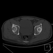
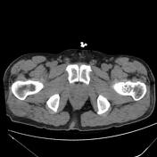
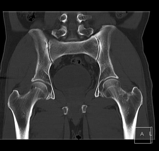
There is an intramedullary lesion measuring 25 x 30 x 35 mm located along the neck of the left femur. This lesion exhibits chondroid matrix calcifications.
There is no evidence of cortical breakthrough or soft tissue involvement.
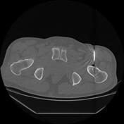

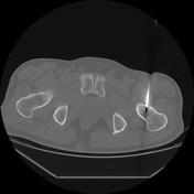
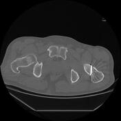
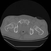
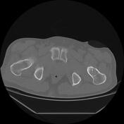

CT guidance was used for the biopsy, performed with sterile techniques. Local anesthetic and IV propofol were administered under the supervision of an anesthesiologist. Two bone specimens were obtained from the left femur, with no immediate post-procedure complications. The patient recovered in the Radiology Department day ward.
Case Discussion
CT image findings suggested differential diagnoses of enchondroma and, less probably, chondrosarcoma. A core needle biopsy was performed, and the histopathology results confirmed the diagnosis of enchondroma.


 Unable to process the form. Check for errors and try again.