Presentation
Right sided lower neck swelling.
Patient Data
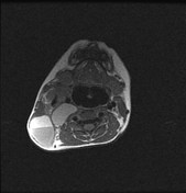


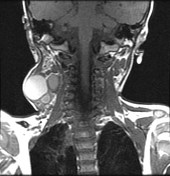

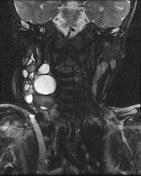

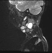

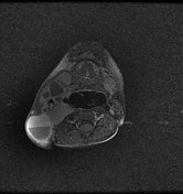

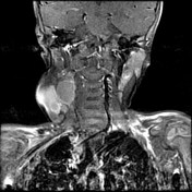

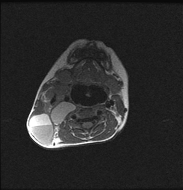
Large multi-locular cystic lesion is noted involving the right posterior cervical space underneath the sternocleidomastoid muscle which is seen elevated. It extends to the carotid sheath, parotid as well as peri-vertebral spaces. It shows mainly intermediate signal in the T1WIs and hyper-intensity signal in the T2WIs. No extension into the mediastinum noted. The related jugular vein is relatively compressed however still patent and the surrounding fat planes are intact. It has multiple sepate and fluid –fluid levels are noted in some of the cysts raise the possibility haemorrhagic complication. Also some of the septae are thick and considerable enhancement in the post contrast T1WIs.
Case Discussion
The described radiological features of well-defined thin-walled cysts with multiple septations and a characteristic midline septum representing the nuchal ligament are matching with Cystic hygroma that also known as cystic or nuchal lymphangioma. It has to be differentiated from an occipital meningocele that has no septations and is seen in direct continuity with a defect in the calvaria.


 Unable to process the form. Check for errors and try again.