
Presentation
Gradual reduced bilateral vision
Patient Data
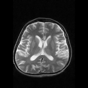

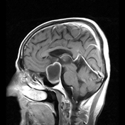
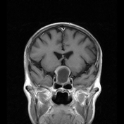

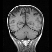
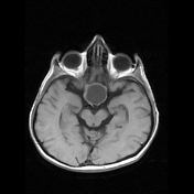
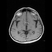

There is a well-defined, altered signals intensity lesion which is involving sellar and supra-sellar regions and is measuring 2.8 x 2.7 x 3.6 cm (AP xWx CC) in size. Returning signals are hypointense on T1 and hyperintense on T2 and T2 FLAIR images with peripheral enhancement on post-contrast images. The lesion is in-separable from pituitary gland and abutting bilateral internal carotid arteries, however flow void is maintained.
The lesion is compressing and displacing the optic chiasm superiorly.
Case Discussion
Findings are of sellar and supra-sellar regions mass lesion likely arising from pituitary gland as described with a differential diagnosis of cystic pituitary adenoma preferred over a Rathke’s cleft cyst.


 Unable to process the form. Check for errors and try again.