
Presentation
Headache and visual field deficit.
Patient Data
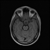


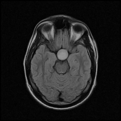
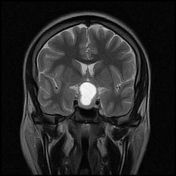
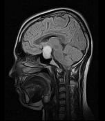
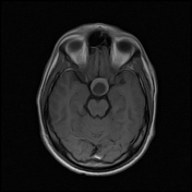
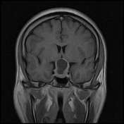

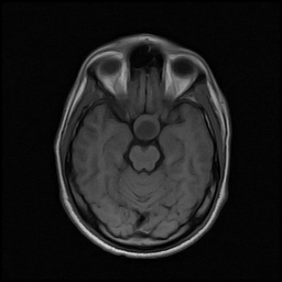
The pituitary gland is enlarged, widens sella turcica, and shows suprasellar extension giving a snowman appearance. It shows a midline intrapituitary cystic lesion with homogeneous low signal intensity on T1 and high signal intensity on T2 and FLAIR. There is diffuse homogeneous post-contrast enhancement of the solid component. It is indenting the optic chiasm and floor of the third ventricle.
Case Discussion
MRI features are in keeping with pituitary macroadenoma with cystic component (cystic pituitary macroadenoma).
For this appearance, the differential diagnosis would be:
Rathke cleft cyst: an intrapituitary cyst with signal change on T2 and surrounded by thin stretched pituitary parenchyma. It can also have a suprasellar extension
pituitary apoplexy: sudden onset of symptoms due to sudden pituitary gland expansion by haemorrhage. MRI signal of the cystic component would reflect haemorrhagic nature (e.g high T1 signal)
adamantinomatous craniopharyngioma: occurs in children with calcifications and irregular soft tissue component
The markedly enlarged and smoothly remodelled pituitary fossa, the excessive amount of soft tissue and the underlying relative frequency of these lesions, makes a pituitary macroadenoma by far the most likely diagnosis.


 Unable to process the form. Check for errors and try again.