
Presentation
Known smoker presents with cough for 3 months.
Patient Data
Age: 60 years
Gender: Male
From the case:
Dense hilum sign
Show annotations
Download
Info
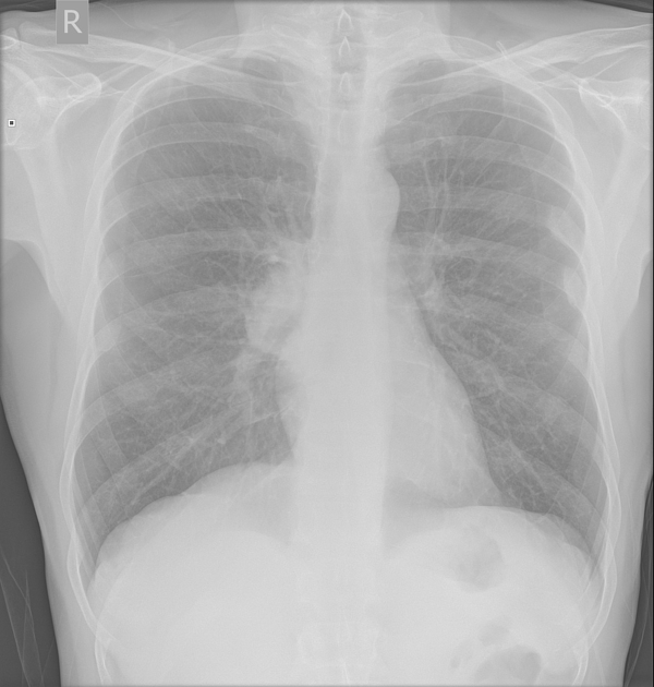
The right hilum is denser than the left.
The normal outline of the right hilar vessels is partly preserved.
From the case:
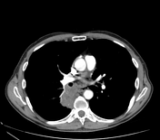
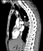
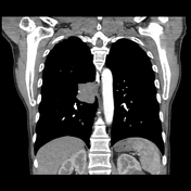
Dense hilum sign

Show annotations
Download
Info

There is a paramediastinal right lower lobe soft tissue mass posterior to the right hilum. On soft tissue windows, the border appears microlobulated.
There are enlarged mediastinal and right hilar lymph nodes.
Case Discussion
This is a histologically confirmed case of non-small cell carcinoma favouring adenocarcinoma.
This case shows the importance of dense hilum sign.


 Unable to process the form. Check for errors and try again.