
Presentation
History of increasing irritability and head circumference. Outside hospital CT demonstrated a right supratentorial mass as well as hydrocephalus.
Patient Data


MRI without & C+ & MR Spectroscopy
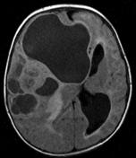
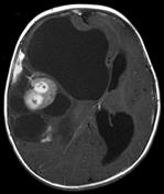
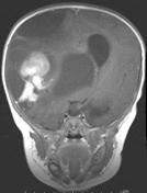
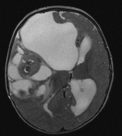
Large tumour that consists of both enhancing solid component and multiple cystic components with marked surrounding oedema, centred within the right frontal lobe.
It causes subfalcine herniation to the left and obstructs the third ventricle at the level of foramen of Monro resulting in marked hydrocephalus. Mass effect on midbrain causing shift of midbrain to the left and uncal herniation.
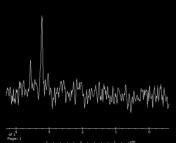
MR spectroscopy obtained from the solid enhancing component of the lesion demonstrates a markedly increased choline peak, consistent with the high cellularity of this mass. An NAA peak is not seen. The solid components of the tumour show restricted diffusion.
The differential diagnosis includes primitive neuroectodermal tumour, atypical rhabdoid/teratoid tumour, anaplastic ependymoma, extraventricular neurocytoma and desmoplastic infantile ganglioglioma.
Case Discussion
Desmoplastic infantile ganglioglioma:
- composed of dural-based desmoplastic stromal tissue and neoplastic astrocytes
- WHO Class 1
- most patients present prior to 2 years of age
- large cystic mass with enhancing nodules and adjacent pial/dural enhancement is characteristic
- solid components are hypodense on T2-weighted images


 Unable to process the form. Check for errors and try again.