
Presentation
Long-standing diabetes, underwent toe amputation.
Patient Data

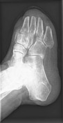
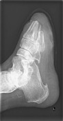
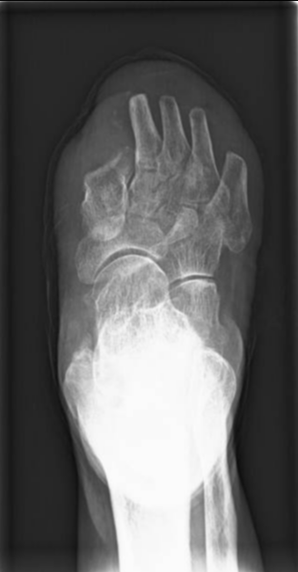
All toes amputated, reaching midshaft of metatarsal bones.
Osteopenic texture of examined bones.
Skin defect related to anterior plantar aspect of 5th metatarsal bone.
Plantar calcaneal spur.
Faint atheromatous plaque along posterior aspect of ankle joint.
No signs of active osteomyelitis.
Maintained ankle joint spaces.
Mild soft tissue swelling around ankle joint.
Case Discussion
Diabetic foot with soft tissue swelling, as well as vascular calcifications.
The complications of diabetes can often be detected radiologically on x-ray foot, including:
- arterial calcification
- soft tissue ulceration, particularly in the lower limbs and feet.
- Charcot joint
- osteomyelitis
So, these are the most important points to look for on diabetic foot plain x-ray.
Although radiographs are important for assessing the position of the bones to each other in general, MRI is the method of choice in establishing an early diagnosis, as well as in monitoring the course of disease activity and in diagnosing infectious complications.


 Unable to process the form. Check for errors and try again.