
Presentation
Diagrams to explain how a paralysed diaphragm can appear normal on fluoroscopy when the patient is erect.
Passive diaphragmatic movement


The weight of the abdominal viscera affects their distribution.
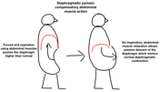
First diagram: how the abdominal muscles compensate for diaphragmatic palsy in the erect position.
In the erect position abdominal muscle contraction in expiration causes diaphragmatic elevation. On inspiration, the abdominal muscles relax and the diaphragm falls mimicking normal diaphragmatic contraction.
Second diagram: how the distribution of abdominal viscera changes in the erect and supine positions.
In the supine position the abdominal viscera are redistributed, flattening the abdomen and pushing upwards against the diaphragm. This limits the ability of the abdominal muscles to force the diaphragm upwards on expiration and limits the subsequent passive fall of the diaphragm on inspiration.
Author: Liz Silverstone.
Case Discussion
These diagrams are intended to demonstrate the importance of recumbent fluoroscopic examination in the assessment of diaphragmatic motion.
In the erect position patients may compensate for diaphragmatic paresis by using abdominal muscle contraction to raise intra-abdominal pressure and force the diaphragm superiorly. On inspiration the abdominal wall muscles relax causing the abdominal viscera to fall so that the diaphragm descends passively, thus mimicking normal diaphragmatic contraction on fluoroscopy.
Fluoroscopy in the recumbent position minimises this effect and is therefore an important part of the routine examination protocol.
The loss of abdominal compensation in the recumbent position also explains why diaphragmatic paresis can cause orthopnoea, most severe when both hemidiaphragms are paralysed.


 Unable to process the form. Check for errors and try again.