
Presentation
Cerebellar signs
Patient Data
Age: 5 years
Gender: Male
From the case:
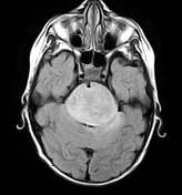
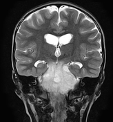
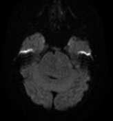
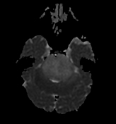
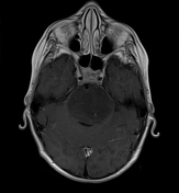
Diffuse brainstem glioma


Download
Info
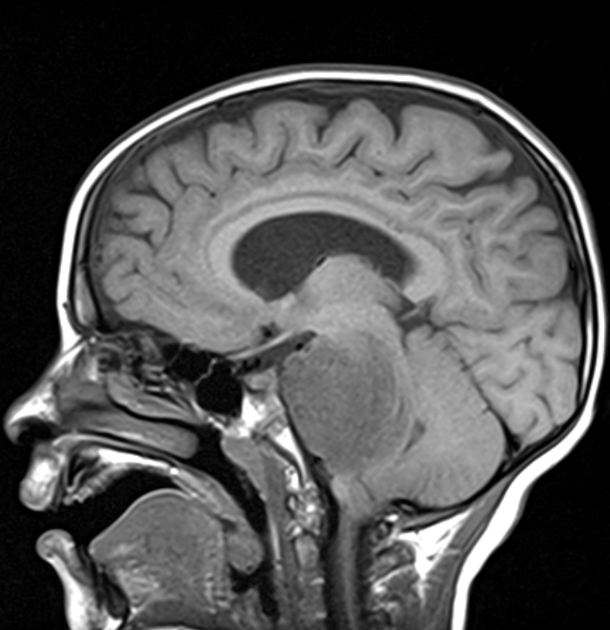
There is a relatively ill-defined mass enlarging the brainstem (centered on the pons), with encasement and anterior displacement of the basilar artery against the clivus, mass effect on the 4th ventricle which is laminated and displaced posteriorly with tonsillar herniation. It is of low signal on T1, heterogeneous high signal on FLAIR/T2 with small punctate areas of enhancement on postcontrast sequences. No restricted diffusion is seen on DWI/ADC. Dilated 3rd and lateral ventricles indicating obstructive hydrocephalus.
Case Discussion
MRI features are most consistent with a diffuse brainstem glioma.


 Unable to process the form. Check for errors and try again.