
From the case:
Diving ranula - through dehiscence


Download
Info

CT of the floor of mouth in a patient with a left-sided swelling demonstrates a large rounded water density cystic mass located inferior to mylohyoid muscle.
Anteriorly a small extension of the cyst can be traced through the muscle fibres into the sublingual space.
Features are consistent with a diving ranula.
From the case:
Diving ranula - through dehiscence
Download
Info
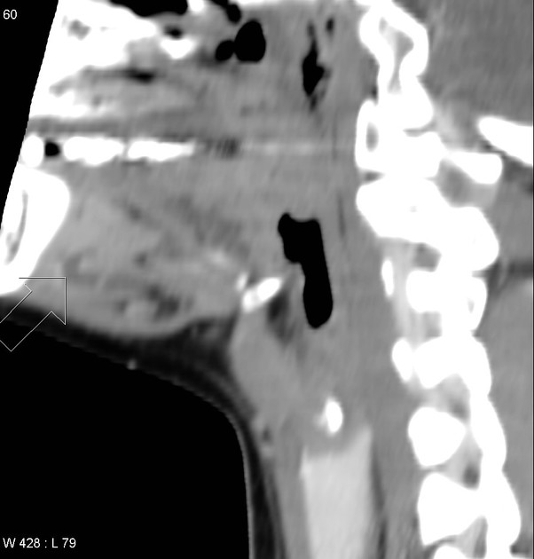
Small extension into the sublingual space is the key to raise the possibility of a diving ranula.
Case Discussion
Final Diagnosis:
Left submandibular gland excision:
- A. Benign granulation tissue and sinus tract with abundant macrophages
- B. Features are consistent with, but not diagnostic of, a plunging ranula
- C. Benign lymph node
- D. Unremarkable submandibular gland


 Unable to process the form. Check for errors and try again.