
Presentation
Bilateral lower limb motor weakness and sciatica. Low back pain.
Patient Data


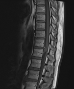
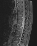
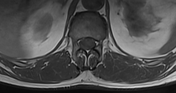
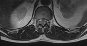
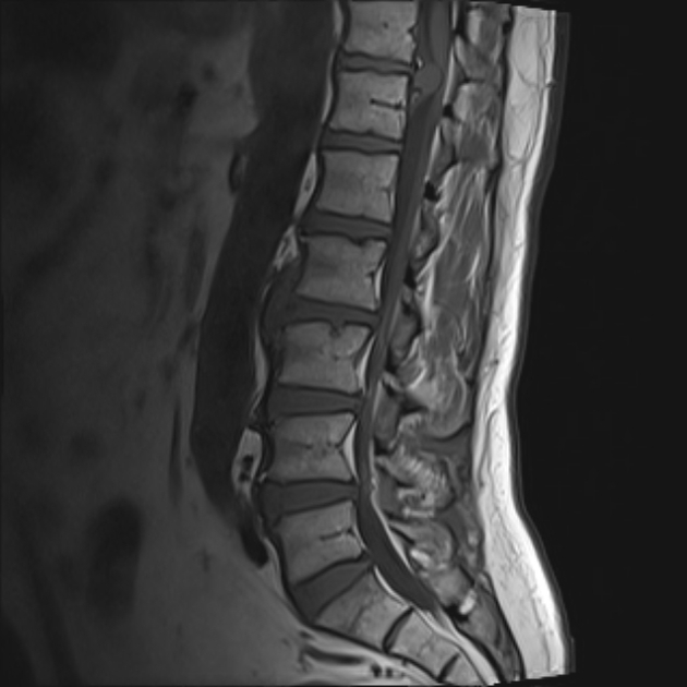
T11/12 focal large posterior midline disc herniation with a narrow neck and broad apex (disc extrusion) and posterior annulus tear showing minimal epidural caudal migration. It is connected to the related disc with no evidence of sequestration. It is markedly compressing the conus medullaris. It elevates the related posterior longitudinal ligament. It is averaging 11x14 mm. No appreciable post-contrast enhancement.
Case Discussion
Imaging features represent midline dorsal central disc extrusion with minimal caudal migration and marked mass effect upon the conus medullaris. Disc extrusion is a type of intervertebral disc herniation and is distinguished from a disc protrusion in that it has a narrow neck and extends above or below the disc level. It is associated with a defect in the annulus fibrosus which allows herniation of nucleus pulposus beyond the confines of the disc. It also shows no evidence of interrupted connection with the original disc and this differentiates it from disc sequestration.


 Unable to process the form. Check for errors and try again.