
Presentation
Cranial nerve VI (CN VI) palsy on physical exam.
Patient Data


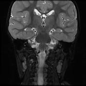
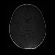
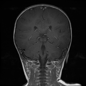
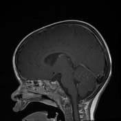
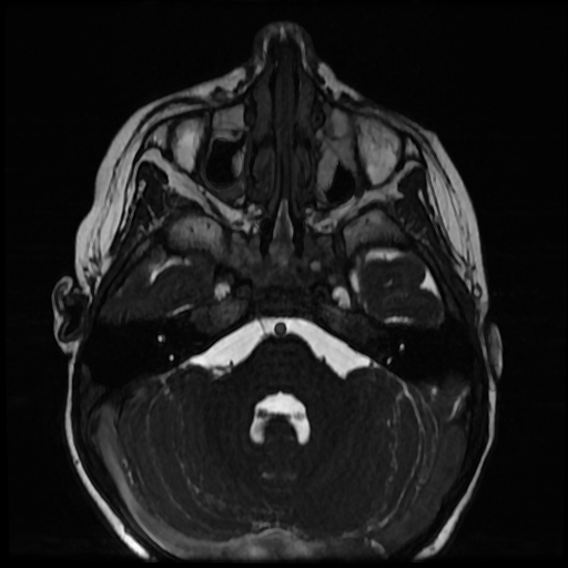
On the high-resolution axial 3-D FIESTA sequence, there is a normal origin of the right CN VI at the pontomedullary junction with a normal cisternal course of the nerve toward the Dorello canal.
A normal left CN VI is not visualized on the left side. There is a thin curvilinear structure arising from the left ventral aspect of the upper medulla, which is below the origin of the contralateral right CN VI. This extends ventrolaterally and appears to split. This could be related to a vessel. Alternatively, this may be aberrant origin of the hypoplastic left CN VI.
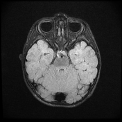
The extraocular muscles are normal in bulk and relatively symmetric. Specifically, there is no significant asymmetry of the lateral rectus muscles to suggest denervation.
Case Discussion
This is a suspected case of Duane syndrome (non-progressive strabismus secondary to an abnormal CN VI). There appears to be an absent CN VI on the left, corresponding to the patient's symptoms.
In this case, the left lateral rectus muscle appears symmetric to the right. Given this, this may indicate that there is incomplete aplasia of the nerve (small non-visualized fires innervating the lateral rectus muscle) or there are aberrant branches of the left CN III supplying the left lateral rectus muscle (Duane syndrome).


 Unable to process the form. Check for errors and try again.