
Presentation
A newly diagnosed cervical cancer patient who presented for a CT scan of the abdomen and pelvis as part of her work-up for disease staging.
Patient Data


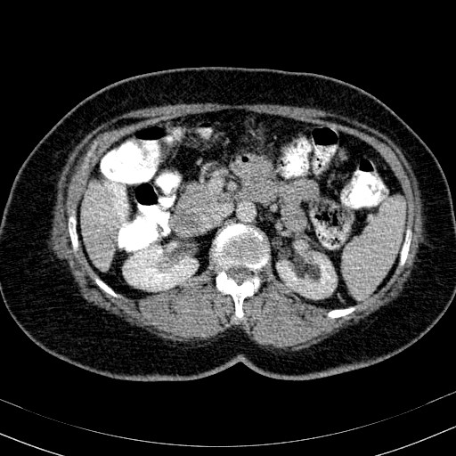
Coronal contrast-enhanced CT images in the arterial phase which demonstrate features consistent with a left-sided duplex renal collecting system, with two renal pelvises an upper and a lower ureteric moiety.
The left-sided ureteric moieties crossover at a level coinciding with the junction of the upper third and middle third parts of the ureters.
The distal attachments/ anatomy of the two left-sided ureteric moieties is unclear, as a delayed (excretory) phase was not performed.
The right kidney and collecting system is normal in appearance, with no duplex system.
Case Discussion
The most common ureteric anomaly is that of ureter number, such as a duplex renal collecting system. The renal parenchyma may be divided into upper and lower components by a septum of Bertin, which is associated with a bifid collecting system and two renal pelvises.
The double ureters may join proximal or distal to the ureteropelvic junction, doing so in the latter case with a 'Y-configuration'. In complete duplication, the upper moiety ureter has a tendency to insert lower (i.e ectopically medially and below) the normal ureteric orifice. The lower moiety inserts orthotopically but tends to reflux (Weigert-Meyer law).
Voiding cystourethrogram (VCUG) or CT intravenous urogram/ pyelogram (CT IVU/IVP) will adequately visualise these systems, their anatomical relations, and any complications such as focal or diffuse hydronephrosis, urine reflux, distal ureteric stricturing and ureterocele formation.


 Unable to process the form. Check for errors and try again.