

Presentation
Headache, right hemiplegia.
Patient Data
Age: 45 years
Gender: Female
From the case:
Dural venous thrombosis


Show annotations
Download
Info
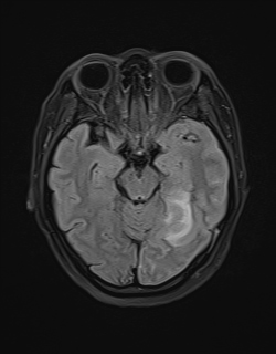
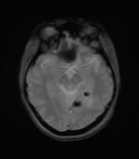
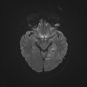
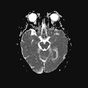
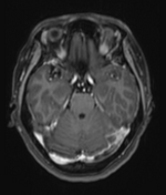
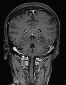
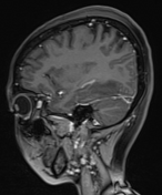
Scattered cerebral haemorrhages in the left temporal region with surrounding brain oedema. The haemorrhagic foci show low signal on GRE, high signal on T1W and T2W, with diffusion restriction.
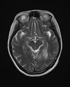
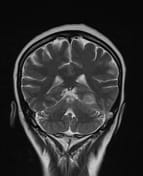
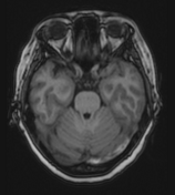
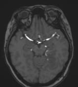
Increased signal intensity on T1W, T2W, and FLAIR in the left transverse sinus, sigmoid sinus, and internal jugular vein; low signal on GRE, with absence of flow on MRV and enhancement on T1FS + GD; findings consistent with venous sinus thrombosis.
No arterial abnormalities detected on MRA.
Case Discussion
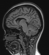
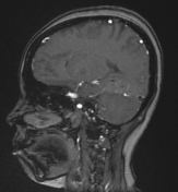
Imaging findings are consistent with dural venous sinus thrombosis causing cerebral venous infarction, manifesting with haemorrhage.
The patient was treated with anticoagulation therapy.


 Unable to process the form. Check for errors and try again.