
Presentation
Cerebellar mass found on trauma CT. Further evaluation using MRI.
Patient Data
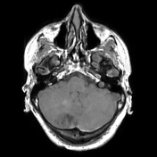

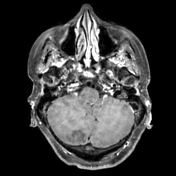

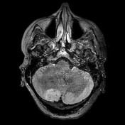
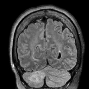

There is a heterogeneous T2 intermediate to hyperintense lesion within the posterior medial aspect of the mid to upper right cerebellar hemisphere demonstrating regional widened cerebellar folia with a tigroid appearance. There is corresponding intermediate hypointense signal in this region on T1-weighted imaging and no significant associated enhancement.
There are smaller satellite lesions anteroinferiorly and superolaterally. Numerous other faint areas of increased T2 signal are seen contralateral, the largest ovoid focus of hyperintense signal is best seen on the coronal FLAIR sequence. These lesions do not display abnormal enhancement.
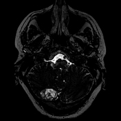
Other findings include an extra-axial, dural-based, homogeneously enhancing mass arising from the ventral aspect of the left tentorial leaflet in the region of the petroclival ligament presumably a meningioma.
Case Discussion
This is a case of a dysplastic cerebellar gangliocytoma and the patient underwent a right suboccipital craniotomy with resection of the tumour.
Histopathology of the mass revealed enlarged, variably-sized ganglion cells that expanded the cerebellar cortex and appeared to replace the granule cell layer. Dystrophic calcifications were present. On immunohistochemistry, the tumour was positive for Neu-N and synaptophysin. The Ki-67 and p53 labellingwere low. These findings were consistent with a cerebellar dysplastic gangliocytoma (Lhermitte-Duclos disease).


 Unable to process the form. Check for errors and try again.