
Presentation
Female presented due to noticeable swelling in her left earlobe
Patient Data
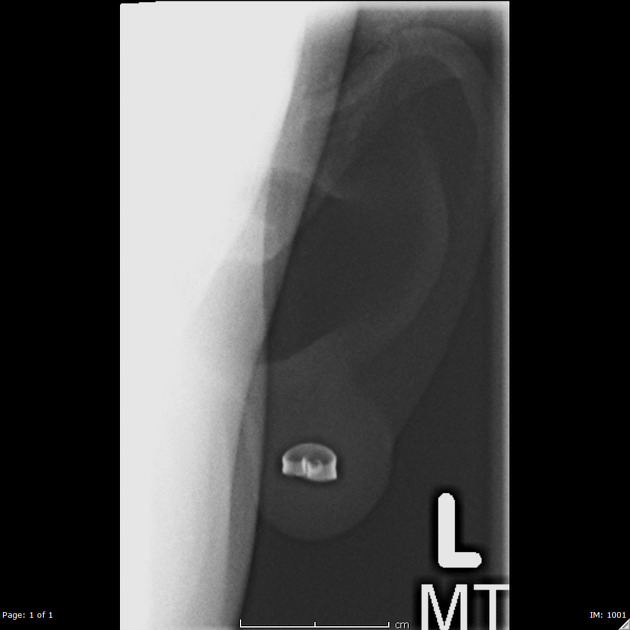
AP view of the Ear/pinna was obtained.
A 7 x 5 mm sized metallic appearing density in the shape of an "earring backing" is seen projecting in the middle of the ear lobule in the expected region of an earring, and is consistent with a retained foreign body. The remainder of the pinna of the ear is intact. No abnormalities or calcifications appreciated.
Case Discussion
Foreign bodies from piercings are a common finding for primary care and emergency physicians. In the pinna, most foreign bodies are from a dislodged earring backing. Most patients experience erythema, inflammation, purulent drainage, and pain on palpation at the piercing site. If the back of the earring is not clearly visible, plain radiographs can be used to confirm the diagnosis. Embedded earrings are commonly found in adolescents under the age of 10 who frequently touch the earring backing. If the foreign body is not removed from the pinna or cartilaginous portion of the ear, an infection and disfigurement may occur. A local anesthetic may be applied to the ear for foreign body removal. If perichondritis or chondritis ensues, antibiotic treatment is appropriate. The ultimate goal is to prevent necrosis and permanent disfigurement.
This case was submitted with supervision and input from:
Soni C Chawla, MD
Associate Professor
Department of Radiological Sciences
David Geffen School of Medicine at UCLA
Olive View - UCLA Medical Center


 Unable to process the form. Check for errors and try again.