
Presentation
The patient presented after a fall down stairs. Imaging was performed as part of trauma workup.
Patient Data
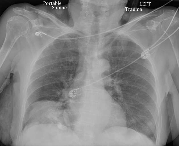
AP radiograph of the chest demonstrates an opacity at the right lung base. The right hemidiaphragm silhouette remains visible as does the right heart border, indicating that this is not within the right lung itself.

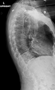
Thoracic spine radiographs (particularly the lateral view) show that the opacity is posterior in the lower chest/upper abdomen.

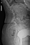
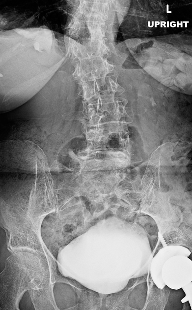
Lumbar spine radiographs (particularly the AP view) show contrast opacifying the mass in the right lower chest/upper abdomen, in a pattern suggestive of a renal pelvis. There is contrast in the urinary bladder. A left total hip arthroplasty is partially imaged.
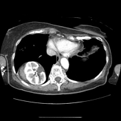


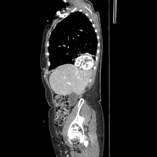
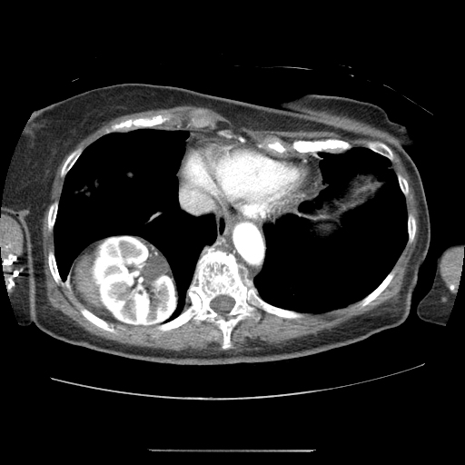
The right kidney is located superior to the liver, projecting into the right thorax. This corresponds to the opacity seen on the radiographs. There is a right posterior diaphragm defect (Bochdalek hernia). There is mild atelectasis at the right lung base. No acute traumatic injury is identified. The left kidney is normal in position.
Case Discussion
Ectopic intrathoracic kidney is an anomaly where one kidney resides in the chest rather than retroperitoneally 1. The etiology of ectopic intrathoracic kidney may be blunt injury, diaphragmatic abnormalities, or congenital developmental anomaly 2. It is typically asymptomatic 3.
In this case, the condition was discovered incidentally on imaging performed as part of a trauma workup. There was a right posterior diaphragm defect (Bochdalek hernia), and these are known to be associated with ectopic thoracic kidneys. No hemorrhage or laceration was seen to suggest a traumatic cause. The condition was asymptomatic in this patient. The right adrenal gland was not clearly visible.
Case co-author: Perry Veras (Loyola University)


 Unable to process the form. Check for errors and try again.