
Presentation
Proptosis
Patient Data


A dense sclerotic lesion seated in the greater wing of the right sphenoid bone and squamous temporal bone with a shaggy margin causing right-side proptosis.

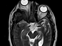


Expanded right sphenoid bone, which appears as a signal void on all pulse sequences significant enhancement in post-contrast study causing right side proptosis.
The smooth meningeal thickening and enhancement seen along the anterior and middle cranial fossa is intimately associated with the right sphenoid bone, resulting in dural tail attachment.
Mild age-related brain atrophies are observed.
Case Discussion
This woman has negative past medical history and complains of slowly progressive right-side proptosis. Surgical operation was done and histopathological result goes with en plaque meningioma.


 Unable to process the form. Check for errors and try again.