
Presentation
Chronic headaches with left exophthalmos. Had surgery of the left orbit 4 years ago
Patient Data
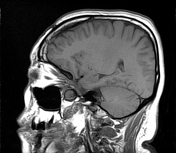

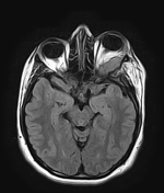
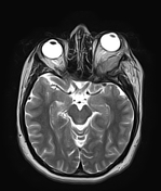
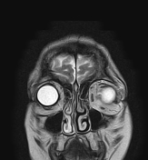
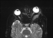
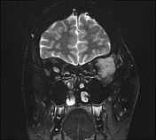

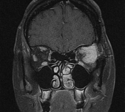

The MRI sequences revealed a well-defined fusiform extraconal soft tissue mass of the superolateral aspect of the left orbit, adjacent to the greater sphenoid wing, isointense on T1WI, and FLAIR, and slightly hyperintense on T2WI with intense and homogeneous enhancement on postcontrast sequences.
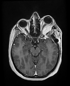
Dural thickening with enhancement of the anterior aspect of the middle cranial fossa extending to the ipsilateral cavernous sinus. The lateral rectus muscle is thickened and displaced medially with non-axial proptosis grade III. The left optic nerve is displaced medially with preserved thickness, and signal intensity.
Bony defects of the greater wing of the sphenoid are noted due to the previous surgery.
Case Discussion
This case illustrates the typical features of en plaque meningioma arising from dura overlying the greater wing of sphenoid with orbital extraconal extension (histologically proven).


 Unable to process the form. Check for errors and try again.