
Presentation
Trauma during a football match. Incidental finding.
Patient Data
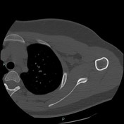

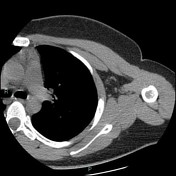
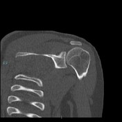
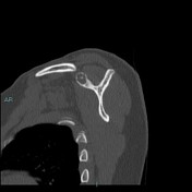

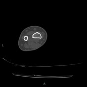
Osteolytic, well-defined, lobulated lesion, in the base of the coracoid process. It contains small calcifications with cortical erosion.
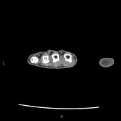
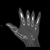
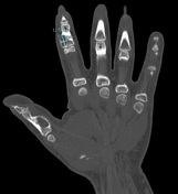
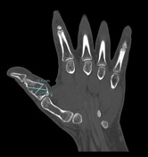
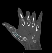

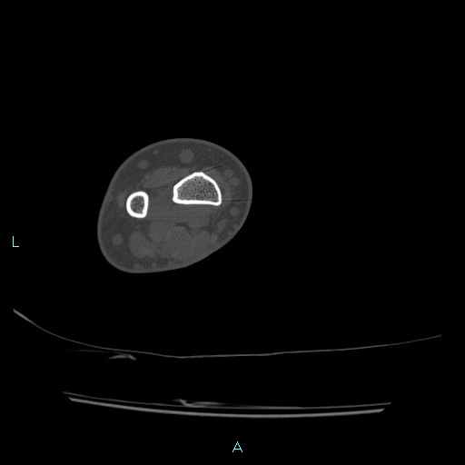
There is an expansile lytic lesion of the 1st metacarpal containing amorphous matrix calcifications characteristic of an enchondroma. There is no pathologic fracture. Another two expansile lytic lesions with internal amorphous calcified matrix are in the proximal phalanx of the thumb and the middle phalanx of the second finger.
Case Discussion
Well-defined lytic and slightly expansile lesions with internal calcification and endosteal thinning in the scapular and hand. Internal calcifications tend to resemble “rings and arcs” of cartilage calcification. Also, these frequently occur in the humerus, femur, tibia and ribs. Rapid growth of lesions or pain not related to a pathologic fracture may suggest malignant transformation. Multiple enchondromas occur in Ollier's disease (enchondromatosis) with a greater incidence of malignant transformation because there are more lesions present.
Radiographer: TSRM Fabio Imola


 Unable to process the form. Check for errors and try again.