
Presentation
Abnormal uterine bleeding after menopause.
Patient Data


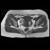
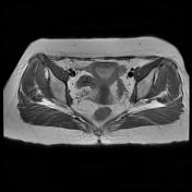
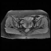
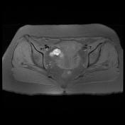
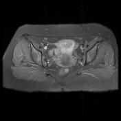

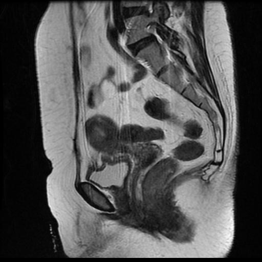
There is a soft tissue mass in the endometrial cavity measuring about 7*11*16 mm demonstrates homogeneous isointense signal intensity on T2 weighted images with myometrial extension.
The uterine cervix and vagina are normal.
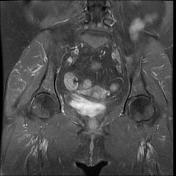
The parametria are clear. Polycystic ovaries.
No pelvic lymphadenopathy or omental lesions.
Both ovaries show atrophic changes with no evidence of solid or cystic adnexal mass lesions .
Case Discussion
The endometrial cavity mass was resected and pathology diagnosis was :
- Endometril intraepithelial neoplasia/ endometrioid carcinoma
- Myometrial invasion can not be excluded.
The recent case is categorized as stage 1a according to FIGO Staging System for Endometrial carcinoma:
- 1a: tumor confined to the body of the uterus with no or less than 50% myometrial invasion


 Unable to process the form. Check for errors and try again.