
Presentation
Presented with headaches and vertigo.
Patient Data


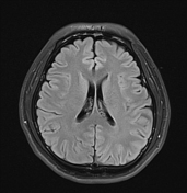
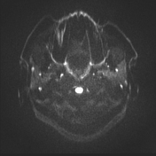
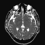
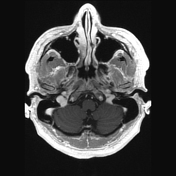
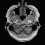
There is an irregular lesion in the right cerebellopontine angle that is partially defined. The lesion is heterogeneous, exhibiting cerebrospinal fluid (CSF) signal intensity on both T1 and T2-weighted images. It shows partial suppression on FLAIR and restricted diffusion on DWI, with an absence of any enhancement in post-contrast T1 imaging. These findings are suggestive of an epidermoid cyst. The lesion extends into the prepontine cistern without causing any mass effect on adjacent structures.
Case Discussion
The epidermoid cyst mimics CSF on MRI, except for DWI which demonstrates restricted diffusion, similar to the study.
Typically patients are between 20 and 40 years of age, as is the case here.
This case demonstrates typical appearances of a large cerebellopontine angle epidermoid cyst.


 Unable to process the form. Check for errors and try again.