
Presentation
Recent onset of headaches, nausea and decreased hearing.
Patient Data
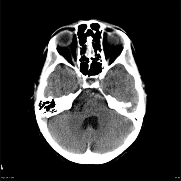
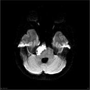
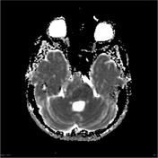
A mass in the right side of the prepontine cistern is largely isodense to CSF but contains some high attenuation strands. It measures approximately 34 x 30 mm in axial diameter and displaces the pons to the left and posteriorly. There is some compression of the fourth ventricle but no dilatation of the lateral or third ventricles. No calcification within the lesion and no hemorrhage.
No intra cerebral lesion elsewhere in the brain. No bone lesion.


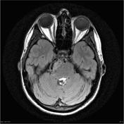
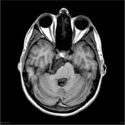
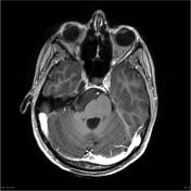
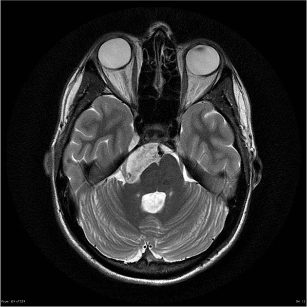
Pre and post contrast scans were performed demonstrating a mass in the right aspect of the pre- pontine cistern that has heterogeneous internal signal with irregular areas of intermediate T2 and FLAIR signal and exhibiting restricted diffusion. The lesion exerts moderate mass effect on the anterior surface of the pons, right cerebral peduncle and right trigeminal nerve. The medial aspect of the lesion is adjacent to the basilar artery and the superior surface abuts the right posterior cerebral artery. The posterior aspect of the lesion comes in proximity to the origin of the seventh and eighth nerves nerves without definite mass effect. There is no definite abnormal enhancement.
This patient went on to have a posterior fossa craniotomy and resection of the tumor.
MICROSCOPIC DESCRIPTION: Specimens show fragments of laminated keratin and segments of thin keratinizing squamous epithelium with an underlying layer of collagen. Laminated keratin with focal calcification abuts this membrane. No appendageal structures are identified. The overall features are of an epidermal cyst. No evidence of tumor is seen in the specimens submitted.
FINAL DIAGNOSIS: Epidermal cyst.
Case Discussion
This case demonstrates typical location and appearances of an epidermoid cyst. These are easily diagnosed on MRI even though on CT their location similarity to CSF can make them challenging.


 Unable to process the form. Check for errors and try again.