
Presentation
Epilepsy
Patient Data


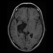
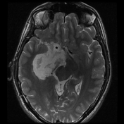
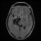
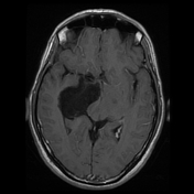
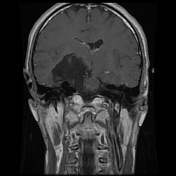
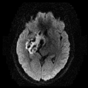
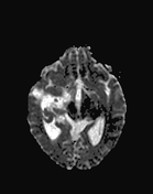
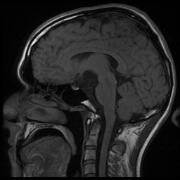
The MRI images reveal an ill-defined lobulated cystic mass of extra-axial location, filling the right cerebellopontine angle, prepontine and ambient cisterns with mass effect on the right middle cerebellar peduncle, 4th ventricle/right lateral ventricle, brainstem, midbrain and right temporal lobe which is compressed and displaced laterally. This cystic mass shows a low signal intensity on T1WI (slightly higher than that of CSF), high signal intensity on T2WI, heterogeneous/dirty signal on FLAIR, and high signal on DWI (restricted diffusion). No enhancement is noted on the postcontrast sequences. Note this mass encases the basilar artery with extension into the ipsilateral internal auditory canal (ICA).
Case Discussion
MRI features characteristic of an intracranial epidermoid cyst.


 Unable to process the form. Check for errors and try again.