
Presentation
Left loin pain. Suspected left renal/ureteric stone.
Patient Data
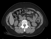

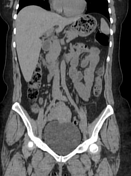
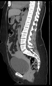
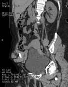

A fat-density ovoid structure adjacent to the descending colon with a thin high-density rim (hyperattenuating ring sign) with surrounding inflammatory fat stranding, and thickening of the adjacent peritoneum.
Case Discussion
When the patient complains of loin pain and the clinician requests a CT-UT investigation in order to exclude renal/ureteric stones; radiologists, particularly junior residents and trainees may only look for stones in the urinary tract. However, it's important to check the whole abdomen and pelvis to exclude other causes of abdominal pain. In this case, it was reported as a normal study by the attending resident who failed to notice the left descending colon epiploic appendagitis!
Therefore, before closing your report on a CT-UT case, check the appendix for early and mild cases of acute appendicitis and move your gaze across the colon to detect subtle cases of epiploic appendagitis!


 Unable to process the form. Check for errors and try again.