
Presentation
Three days history of left lower pelvic pain.
Patient Data
Age: 55 years
Gender: Female
From the case:
Epiploic appendagitis - sigmoid colon
Download
Info
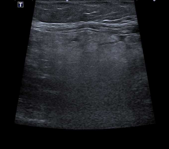
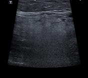
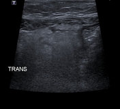
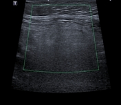
There is an ill-defined non-compressible, hyperechoic mass (4 x 3.5 cm) adjacent to the wall of the sigmoid colon, with no internal vascularity on colour Doppler.
From the case:
Epiploic appendagitis - sigmoid colon

Download
Info
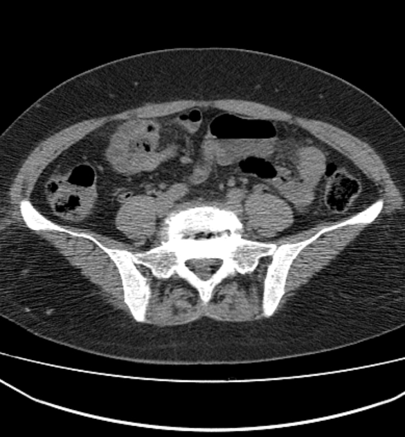
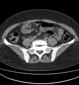
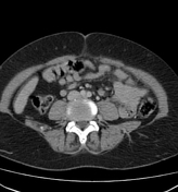
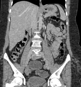
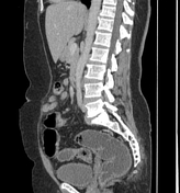
There is an ovoid structure of fat-density, in the left iliac fossa, adjacent to the sigmoid colon with thin peripheral hyperdense rim ( hyperattenuating ring sign) and surrounding inflammatory fat stranding. Minimal and regular thickening of the adjacent sigmoid wall is noted.
A Riedel lobe is noted.
Case Discussion
Ultrasound and CT features of an epiploic appendagitis


 Unable to process the form. Check for errors and try again.