
Presentation
Bilateral proptosis, more in left eye. No previous medical illnesses.
Patient Data


There is diffuse almost symmetrical osteosclerosis with resultant loss of the corticomedullary differentiation mainly in the tibia.


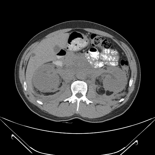
There is diffuse retroperitoneal soft tissue infiltrating mass lesion, associated with bilateral perirenal soft tissue thickening. There is very minimal subcapsular renal fluid and stranding of the Gerota's fascia fat plane giving the appearance of hairy kidneys. The thickening mainly involves the major retroperitoneal vessels such as the superior mesenteric, renal vessels, and inferior mesenteric artery. It also surrounds the abdominal aorta. The thickening extends to surround the mesenteric branches of the superior mesenteric artery as well as the root of mesentery. However, despite the vascular encasement, there is no intraluminal filling defect.
Note: multiple hepatic cysts.
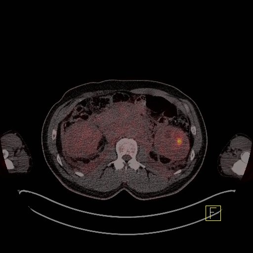
Retroperitoneal and perirenal fibrosis also demonstrating heterogeneous mild increased metabolic activity.


Axial T1 and T2 weighted MR images of the orbits show bilateral hypointense intraconal infiltrating soft tissue masses splaying the extraocular muscles and resulting in proptosis. Fat-suppressed T1-weighted MR image shows diffuse enhancement of the masses.

CT guided biopsy was done by 18 G x 15 cm needle from the left retroperitoneum.
Pathology showed :
- Fibrosis interspersed by foamy macrophages highly suggestive of Erdheim-Chester disease.
- No malignancy is seen.
Case Discussion
Erdheim-Chester disease is a rare and challenging case to diagnose. It shows multi-systemic/organic involvement. Therefore, diagnosis is based on histological and radiological findings.
As in this case, there are multiple findings involving the orbit, manifested by bilateral intraconal soft tissue mass lesions, retroperitoneal or perirenal infiltrative soft tissue thickening and bony sclerosis. When the radiological findings are taken separately, there will be many differential diagnoses.
Lymphoma will be on top of the differential diagnosis because it contains most of these radiological findings.
Additional Contributors:
Dr. Asem Mansour, Consultant Radiologist. Chief Executive Officer/Director General, King Hussein Cancer Center(KHCC), Amman, Jordan.
Dr. Hussain Ali Aby Ali, Consultant Radiologist, Tawam hospital, United Arab Emirates.


 Unable to process the form. Check for errors and try again.