Presentation
Atraumatic pain, swelling of left thigh since 2 months. Reduced range of motion of the knee.
Patient Data
Age: 16 years
Gender: Male
From the case:
Ewing sarcoma - femur
Download
Info
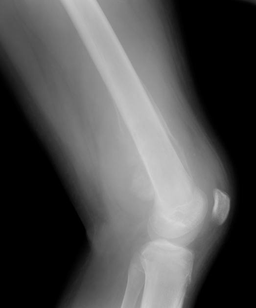
There is a lesion located in the distal femur with typical aggressive features of a malignant tumor with:
- irregular and partial destruction of cortical bone
- periosteal reaction with Codman triangle formation
- indistinct zone of transition
- perpendicular spiculations producing a sunburst appearance
- associated large soft tissue swelling
Histologically proven Ewing sarcoma.
From the case:
Ewing sarcoma - femur


Download
Info
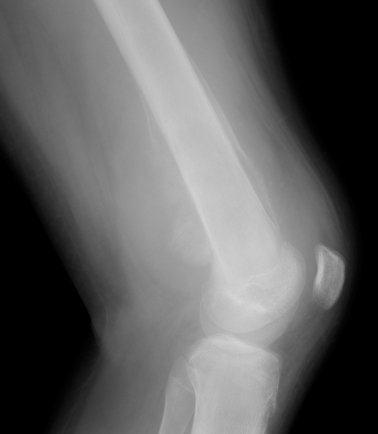
- Green: Cortical bone destruction
- Pink: Periosteal reaction
- Arrow: Codman triangle
- Blue: Sunburst appearance
- Yellow lines: Cortical thinning
- Gray lines: soft tissue expansion
Case Discussion
Pathologically proven Ewing sarcoma. This is a highly malignant bone tumor seen in the first two decades of life. The main differential diagnosis is osteosarcoma.