
Presentation
Increasing swelling and lump at the lateral aspect of right orbital rim for 6 months.
Patient Data


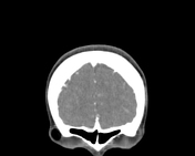
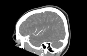
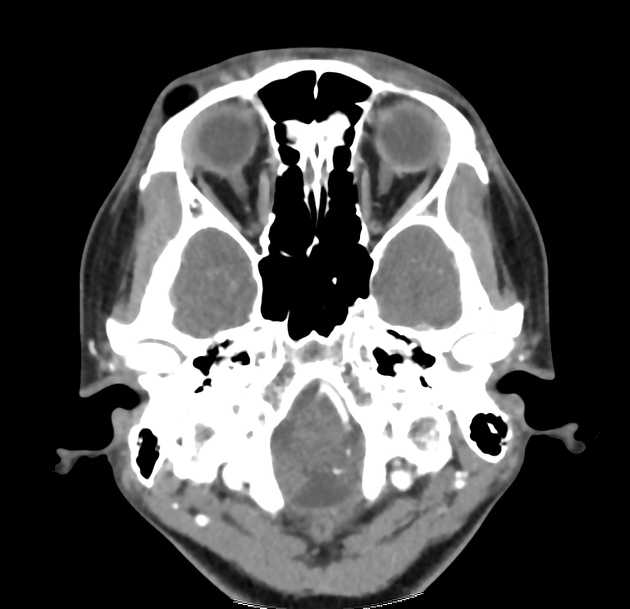
A well-defined subcutaneous lesion at the superolateral aspect of right orbital rim. The lesion is predominantly fat component without significant enhancement post contrast administration. No calcification within. No intraorbital extension. No erosion of the adjacent bone.
Case Discussion
This patient has history of excision of orbital dermoid cyst at the same location of the current swelling 14 years ago. The current mass has been proven to be recurrence of orbital dermoid cyst from the excision.
External angular dermoid is congenital swellings occurring at the outer corner of the eye.
They are usually arising from sites of the lines of embryonic fusion where ectodermal elements fail to disconnect from the developing neural tube or get trapped beneath the skin.
Incomplete excision of orbital dermoid cyst will result in increased rate of recurrence 1. It is advocated that early excision of the orbital dermoid cyst will reduce the risk of recurrence as it is easier for complete excision without disruption of the cyst wall 1.


 Unable to process the form. Check for errors and try again.