
Presentation
Left-sided periorbital swelling along with blurred vision for 3 weeks.
Patient Data
Age: 20 years
Gender: Female
From the case:
External angular dermoid cyst

Show annotations
Download
Info

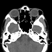
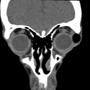
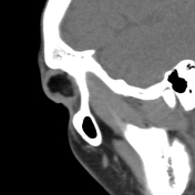
A well-defined fat density mass lesion with mean density of about -134 HU, is noted involving the lateral margin of the left orbit, medially the mass lesion is abutting globe and lateral rectus muscle without any CT detectable invasion. The overlying skin appears intact. No bony destruction is noted.
Right globe and orbit appear normal.
Imaged brain parenchyma appears unremarkable without intra-cranial extension.
Case Discussion
The age of the patient and location of the lesion with imaging findings are more in favor of dermoid cyst as detailed.
Co-contributor Dr, Anwar-ul-haq Zadaran.


 Unable to process the form. Check for errors and try again.