
Presentation
Long-standing painless swelling in the right periorbital region.
Patient Data
Age: 15 years
Gender: Female
From the case:
External angular dermoid cyst


Show annotations
Download
Info
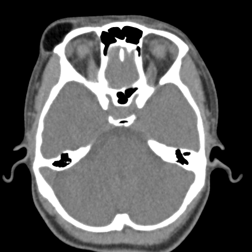
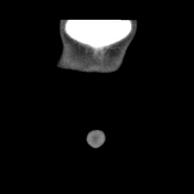
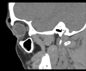
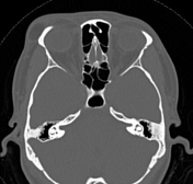
A definable, fat density (-150 HU) lesion is noted in the subcutaneous tissues of the right external angular region.
No evidence of calcification. No intraorbital or intracranial extension is seen.
A small depression in the underlying frontal bone is seen as likely suggestive of remodeling due to the chronicity of the lesion. No widening of the adjacent right fronto-zygomatic suture.
Case Discussion
CT features of the above-mentioned lesion are more in favor of superficial external angular dermoid cyst.
Co-contributor: Dr. Anwar-ul-Haq Zadran.


 Unable to process the form. Check for errors and try again.