
Presentation
The patient presented with discomfort in the left ear. Upon examination, swelling was noted around the ear area.
Patient Data
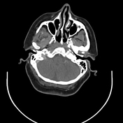

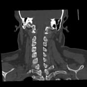

A contrast-enhanced CT scan of the neck reveals significant enlargement of the left parotid gland, exhibiting increased attenuation. This is accompanied by reactive adenopathy in levels 2 and 3, strongly suggesting a diagnosis of parotitis.
Fatty replacement of the right parotid gland with hypertrophy of the right accessory parotid gland.
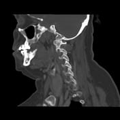
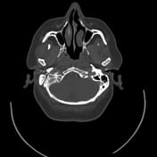
Moreover, the scan shows bilateral symmetrical calcifications in the cartilaginous part of the external auditory canal (EAC).
No calcifications were noted in the external auricle. These findings warrant close attention and may require further evaluation to ensure appropriate management.
Case Discussion
The cause of the patient's ear discomfort was proposed to be inflammation of the LT parotid gland.
The bilateral symmetrical calcification of the external auditory canal's outer cartilaginous part was an incidental finding. The fact that there is no associated calcification of the external auricle makes the case more interesting than the combined calcification of the external auricle and EAC.


 Unable to process the form. Check for errors and try again.