
Presentation
Witnessed collapse striking head. New seizures. Now GCS 9. Left temporal bruising.
Patient Data

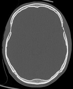
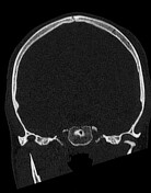

Native unenhanced head, with axial and coronal brain and bone reconstructions.
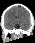
Fracture through the left temporal bone, passing through the lateral canal of the middle meningeal artery, and continuing into the roof and lateral wall of the orbit.
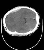
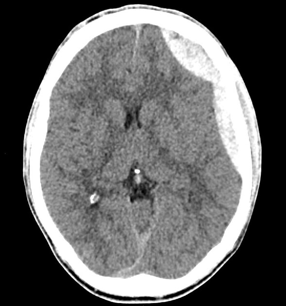
Resultant extradural haematoma - maximal coronal depth 9 mm. Local sulcal effacement but no significant mass effect.
Left orbital roof haematoma with early proptosis.


Following transfer to tertiary neurosurgical centre, further clinical deterioration.
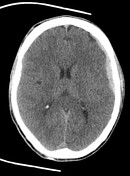
Expansion of the left EDH, including a large newer frontal component, now 19 mm in depth.
Progressive mass effect with partial effacement of the left lateral ventricle, early subfalcine and left uncal herniation.
Case Discussion
The middle meningeal artery is the source of this left extradural haematoma.
The short interval imaging demonstrates the rapid progression of these traumatic insults, and why these cases are neurosurgical emergencies.
The case was managed with immediate decompressive craniotomy and haematoma evacuation.


 Unable to process the form. Check for errors and try again.