
Presentation
Fall onto hard surface. Headache.
Patient Data


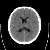
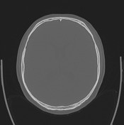
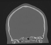
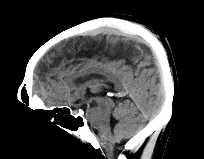
Overlying the parietal bone towards the vertex immediately underneath the parietal bone, there is an area of extra-axial hyperdensity, which is best appreciated on the sagittal reconstructions. There is adjacent subgaleal hematoma overlying this extending posteriorly towards the parieto-occipital bone, but no underlying fracture is seen. There is compression and displacement of the adjacent superior sagittal sinus due to its mass effect.
Conclusion:
Appearances are characteristic of a venous epidural hematoma.
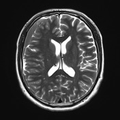
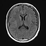
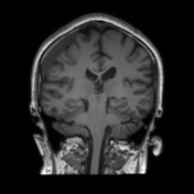
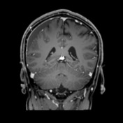
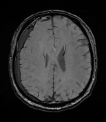

MRI one month later demonstrates a persisting subdural hematoma. The previously demonstrated venous extradural hematoma has resolved.
Case Discussion
The rule of "extradural hematomas shall not cross suture lines" is only valid if the sutures are intact. Even a diastasis which is difficult to appreciate can tear the parietal dura and allow for extradural venous hemorrhage.


 Unable to process the form. Check for errors and try again.