Extraventricular neurocytoma
Diagnosis certain
Updates to Case Attributes
Body
was changed:
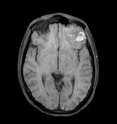
Differential diagnosis in this case would include the following entities:
- Anaplastic oligodendroglioma. The principal differential diagnosis, as this lesion can also manifest with multiple calcifications, bone remodeling, and lack of perilesional edema. On the other hand, this type of neoplasm tends to have coarse and larger calcifications and it is typically encountered in middle-aged adults.
- Chondrosarcoma. The presence of rings and arcs calcifications and bone destruction raised the possibility of chondrosarcoma. Also, the T2 hyperintense regions resembling the cartilaginous matrix favored this option. However, chondrosarcoma does not usually appear as an intracranial mass and is rare in young patients.
- Grade III meningioma. Cystic components are less frequent in this type of lesion, and enhancement is usually more significant and homogeneous.
- Gangliocytoma/ganglioglioma. Ordinarily seen in children and young adults, these tumors can have a similar clinical presentation. Nevertheless, they have a more homogeneous radiological appearance and do not show bone resorption.
- Anaplastic astrocytoma / GBM. These neoplasms rarely show calcifications and bone resorption, and frequently present with marked peripheral FLAIR hyperintensity.
- Metastases. Although this diagnosis should always be considered, there was no oncological history in this patient.
The first diagnostic possibility was a chondrosarcoma, especially due to the T1 and T2 hyperintense components and the rings and arcs calcifications. The patient went to have a surgical resection of the lesion and the histologic examination is showed above. No chondroid matrix was found so a chondrosarcoma was discarded. There were also no
-</ul>- +</ul><p>The first diagnostic possibility was a chondrosarcoma, especially due to the T1 and T2 hyperintense components and the rings and arcs calcifications. The patient went to have a surgical resection of the lesion and the histologic examination is showed above. No chondroid matrix was found so a chondrosarcoma was discarded. There were also no </p>
Updates to Study Attributes
Images Changes:
Image MRI (T1 fat sat) ( update )

Specifics
changed from T1 fat sat to T1 fat sat.
Image 3 MRI (T1 fat sat) ( update )
Position
was set to
.