
Presentation
A hypertensive male patient presented with dizziness, vomiting, left facial palsy, ophthalmoplegia, right upper motor neurone lesion, mild ptosis, diplopia, and nystagmus.
Patient Data


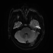
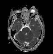
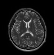
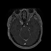
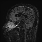

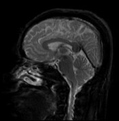
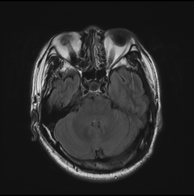
A well-defined inverted comma-shaped focus of restricted diffusion is seen in the most dorsal aspect of the pons off right to the midline in the presumed site of the right facial nerve colliculus in the anatomic elevation of the dorsal pons intimately related to the 6th cranial nerve nucleus and motor fibre of the facial nerve that loop over the abducent nucleus. It appears bright on FLAIR T2 and iso signal on T1WI. No enhancement in the post-contract study or mass effect on the 4th ventricle. Few scattered foci of high signal on FLAIR T2 are seen within the periventricular white matter with no enhancement, blooming, or restricted diffusion. They represent ischaemic strokes.
Case Discussion
The facial colliculus is an elevation on the floor of the fourth ventricle and is not formed by the facial nucleus, but by the fibres of the facial nerve arching backward around the abducent nucleus before turning forwards once more in the caudal pons. So the lesion involving the facial colliculus may result in facial colliculus syndrome by involving: the abducens nerve (CN VI) nucleus, facial nerve (CN VII) fibres at the genu, and medial longitudinal fasciculus.


 Unable to process the form. Check for errors and try again.