
Presentation
Headache and depression.
Patient Data


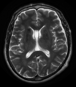
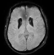
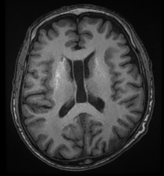
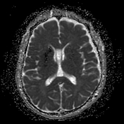
Bilateral basal ganglia abnormal signal which elicits high signal on T1, and low signal on T2, FLAIR, and DWI. On SWI, there is evidence of blooming of the basal ganglia, and to a lesser extent, dentate nuclei.
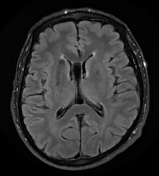
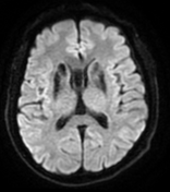
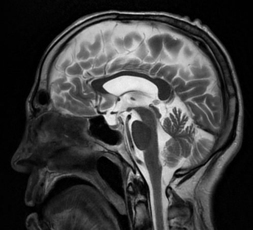
Cavum septum pellucidum et vergae is noted incidentally.
Findings are suggestive of Fahr disease.
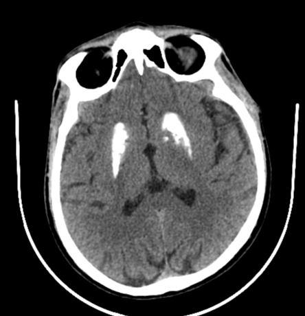
Calcifications are prominent in the basal ganglia, centrum semiovale, and cerebellar hemispheres.
Cavum septum pellucidum et vergae is noted incidentally.
Case Discussion
In this case, the signal changes at the basal ganglia which elicits high T1 signal and low signal on the rest of the sequences were suggestive of calcifications which is confirmed on susceptibility weighted imaging with characteristic signal loss and blooming.
It's very important to remember that the MRI appearance varies depending on the degree of calcification and the stage of the disease and can be remarkably normal in appearance even when CT changes are profound, unless susceptibility weighted imaging is obtained.
Fahr syndrome, also known as bilateral striatopallidodentate calcinosis, is characterised by abnormal vascular calcium deposition, particularly in the basal ganglia, cerebellar dentate nuclei, and white matter, with subsequent atrophy.
It can be either primary (usually autosomal dominant) or secondary to a large number of underlying illnesses or metabolic disturbances.


 Unable to process the form. Check for errors and try again.