Fahr syndrome
Updates to Case Attributes
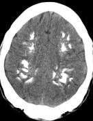
CT brain which easily detects calcium, is the preferred method of localizing and assessing the extent of cerebral calcifications. Most frequently the the calcification is in lenticular lenticular nucleus, especially the internal globus pallidus. Calcifications in the putamen, thalami, caudate, and dentate nuclei are common. Occasionally, calcifications begin or predominate in regions outside the basal ganglia.
Calcification seems to be progressive, since calcifications are generally more extensive in older individuals and an increase in calcification can sometimes be documented on follow-up of affected subjects.
The calcifications in FIBGC areprimary Fahr syndrome are not distinguishable from those secondary to hypoparathyroidism or other causes. Cerebellar gyri, the brain stem, centrum semiovale, and subcortical white matter may also be affected.
-<p><strong>CT brain </strong>which easily detects calcium, is the preferred method of localizing and assessing the extent of cerebral calcifications. Most frequently the calcification is in lenticular nucleus, especially the internal globus pallidus. Calcifications in the putamen, thalami, caudate, and dentate nuclei are common. Occasionally, calcifications begin or predominate in regions outside the basal ganglia.</p><p>Calcification seems to be progressive, since calcifications are generally more extensive in older individuals and an increase in calcification can sometimes be documented on follow-up of affected subjects.</p><p>The calcifications in FIBGC are not distinguishable from those secondary to hypoparathyroidism or other causes. Cerebellar gyri, the brain stem, centrum semiovale, and subcortical white matter may also be affected.</p>- +<p><strong>CT brain </strong>which easily detects calcium, is the preferred method of localizing and assessing the extent of cerebral calcifications. Most frequently the calcification is in lenticular nucleus, especially the internal globus pallidus. Calcifications in the putamen, thalami, caudate, and dentate nuclei are common. Occasionally, calcifications begin or predominate in regions outside the basal ganglia.</p><p>Calcification seems to be progressive, since calcifications are generally more extensive in older individuals and an increase in calcification can sometimes be documented on follow-up of affected subjects.</p><p>The calcifications in primary <a title="Fahr syndrome" href="/articles/fahr-syndrome-1">Fahr syndrome</a> are not distinguishable from those secondary to hypoparathyroidism or other causes. Cerebellar gyri, the brain stem, centrum semiovale, and subcortical white matter may also be affected.</p>
Updates to Study Attributes
Bilateral symmetricalNoncontrast CT shows bilateral symmetrical dense calcific foci in the dentate nucleusnuclei, basal ganglia, thalamus and coronathalami, and corona radiata.
Image CT (non-contrast) ( update )

Image CT (non-contrast) ( update )
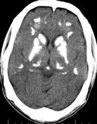
Image CT (non-contrast) ( update )
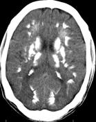
Image CT (non-contrast) ( update )