
Presentation
Shortness of breath 3 days after motor vehicle collision.
Patient Data

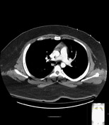
Lungs hypoinflated but clear.


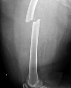
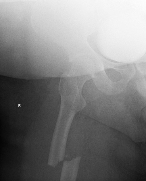
Comminuted fracture proximal shaft right femur with one shaft width medial and posterior displacement.
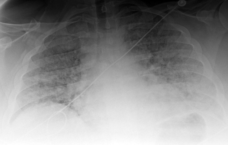
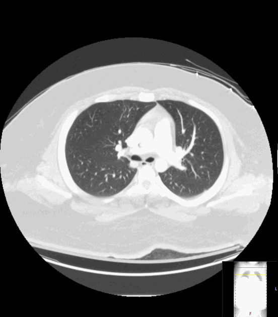
New diffuse increased opacity both lungs.


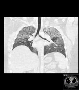
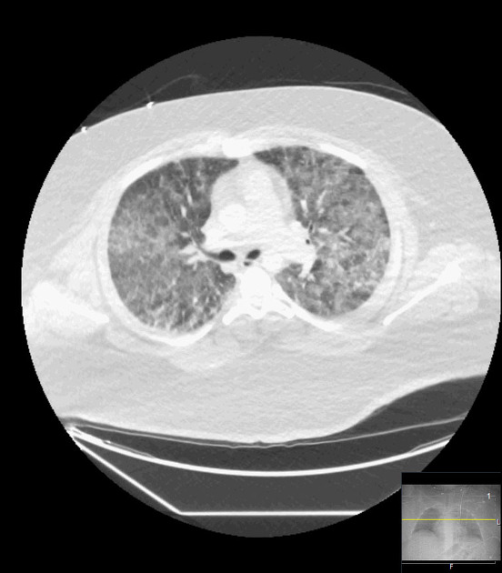
New diffuse bilateral groundglass opacity both lungs, no pleural effusions.
Case Discussion
The ground glass opacity that developed in the lungs 3 days after presentation is non-specific and could be secondary to edema or hemorrhage, but in a patient with a long bone fracture, fat embolism syndrome, while rare, should also be considered. There are 3 major criteria for fat embolism syndrome. Lung involvement characteristically results in shortness of breath and groundglass opacity on CT. Fat emboli may also involve the brain, resulting in mental status changes ranging from confusion to coma. Skin involvement results in a petechial rash.


 Unable to process the form. Check for errors and try again.