
Presentation
Line placement.
Patient Data
Age: 80 years
Gender: Male
Show annotations
Download
Info
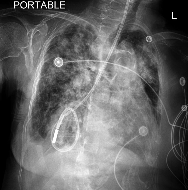
Coiled distal feeding tube projects over the medial right lung base. In the neck, the proximal feeding tube is adjacent to but not in the airway. Enlarged cardiac silhouette, multifocal consolidation.
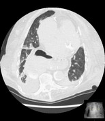
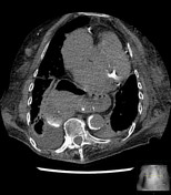
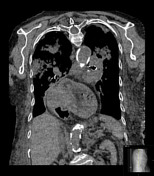
CT chest from one day prior

Download
Info

Large hiatal hernia, multifocal consolidation, small bilateral pleural effusions.
Case Discussion
On the radiograph, the feeding tube tip projects over the medial right lung base, mimicking pulmonary tube placement. But in the neck, the tube is clearly adjacent to and not in the airway. A CT from the previous day shows a large hiatal hernia with a component located just lateral to the right heart border, corresponding to the location of the feeding tube on the next day's radiograph.


 Unable to process the form. Check for errors and try again.