
Presentation
Incidental findings
Patient Data
Note: This case has been tagged as "legacy" as it no longer meets image preparation and/or other case publication guidelines.
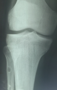
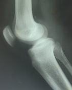
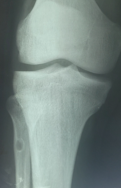
Two radiographic views reveal a small, nearly oval, well-defined cortical lucent lesion located at the junction of the proximal dia-metaphysis of the right fibula. The lesion displays a sclerotic rim and has a narrow transitional zone. There are no obvious signs of periosteal reaction, abnormal soft tissue, or calcification present.
Case Discussion
A fibrous cortical defect is a lesion with the same characteristics and histopathology as non-ossifying fibroma but with a smaller size (< 2cm) than the latter. In the last issue of the WHO classification of soft tissue and bone tumor (5th edition) 1 those lesions have been summarized as non-ossifying fibromas. The term fibrous cortical defect is now discouraged, as is the quality of radiographs like the one above.
It shares the letter "F" with fibrous dysplasia in the popular mnemonic FEGNOMASHIC. This condition is asymptomatic and self-limiting, typically healing completely by adulthood. It is one of the "leave me alone" lesions.


 Unable to process the form. Check for errors and try again.