
Presentation
About 4 years prior to the presentation, the patient had a small laceration on the forehead region due to a road traffic accident. Since then the patient was able to palpate a tiny nodule at the site of injury.
Patient Data
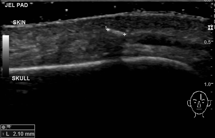
There is a linear echogenic focus (2.1 x 1.2 mm) in the forehead superficial connective tissue. It is a foreign body. There is no surrounding granuloma formation. An adjacent vein is patent. There is no bone cortex irregularity.

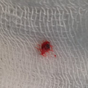
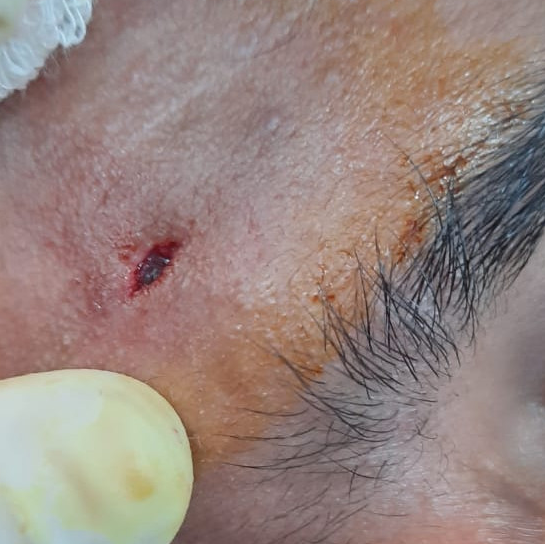
An intraoperative photo shows a small incision and dark-colored foreign body in the soft tissue. 2nd photo shows a retrieved foreign body with a blood clot around it.
Case Discussion
The case shows post traumatic foreign body implantation in the forehead region. The surgical exploration revealed a piece of gravel which was expected by the patient as well as the doctor.
Photos Courtesy: Operating surgeon Dr. Niraj I. Patel.


 Unable to process the form. Check for errors and try again.