
Presentation
Left sided weakness
Patient Data
Age: 70 years
Gender: Male
From the case:
Foville syndrome

Download
Info

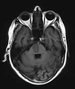
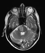
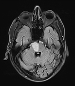
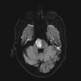
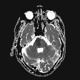
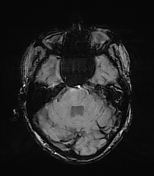
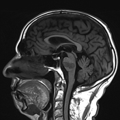
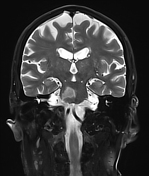
Subacute infarct with diffusion restriction is seen involving inferomedial aspect of pons on right side.
Gliotic area with chronic haemorrhage are seen involving right lentiform nucleus, external capsule.
Lacunar infarcts and areas of ischaemic demyelination are seen in bilateral periventricular, deep and subcortical white matter.
Convexity sulci, lateral ventricles and surface of cerebellar folia are prominent , likely age related atrophy.
Case Discussion
Subacute infarct involving inferomedial aspect of pons on right side consistent with Foville syndrome.


 Unable to process the form. Check for errors and try again.