
From the case:
Frontal nerve (illustration)
Download
Info
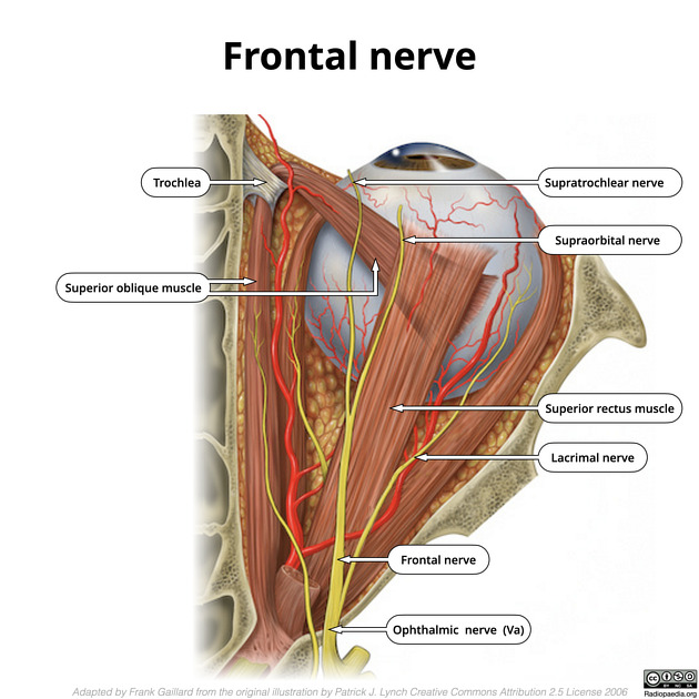
Illustration depicting the frontal nerve and branches.
Note: the supratrochlear nerve actually passes more medial than shown in this illustration, closer to (in fact above) the trochlear pulley.
Case Discussion
This image is an edited and labeled version of an illustration by Patrick Lynch (original Wikimedia commons here).
Changes include:
- moving the origin of the lacrimal nerve posteriorly (it divides before the orbital apex)
- removing a muscular branch from the oculomotor over the lateral rectus (for clarity)


 Unable to process the form. Check for errors and try again.