
Presentation
Mild left side proptosis and hypoglobus and a history of long-standing pansinusitis.
Patient Data


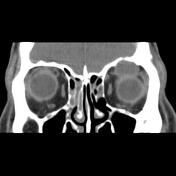

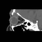
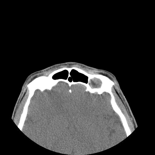
Lytic expansile lesion coronal width up to 22 x 18 mm and axial width up to 20 mm in left frontal sinus extended within left orbital cavity upper segment through orbital roof bone defect with impression on adjacent superior rectus muscle and eye is seen which has caused proptosis and hypoglobus. Mucosal thickening and fluid in all paranasal sinuses are seen.
Case Discussion
The case illustrates the non-contrast MDCT feature of a pathology-proved frontal sinus mucocele. One of the major complications of chronic sinusitis is mucocele formation in paranasal sinuses and the frontal sinus is the most common location that can lead to hypoglobus and proptosis.


 Unable to process the form. Check for errors and try again.