
Presentation
Rhinorrhoea and long-standing headache.
Patient Data
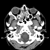


Complete opacification of the frontal sinuses, ethmoidal cells and right maxillary sinus.
Mucous retention cysts in the left maxillary sinus.
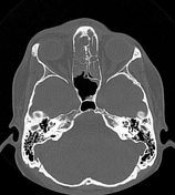
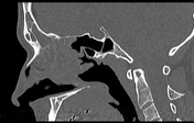
The right maxillary sinus is enlarged and occupies part of the right nasal cavity. A poorly defined area with increased density is observed centrally.
The walls of the right maxillary sinus are thinned and show signs of erosion.
A nonspecific collection is identified in the right orbital cavity's upper and medial border.
Case Discussion
The complete opacification of the right maxillary sinus, with increased size and content with hyperdense elements, is the key to suggesting the diagnosis of fungal sinusitis. The right orbital collection could be an element of invasive sinusitis. Any history of immunocompromise is unknown.
Fungal sinusitis is divided into invasive and noninvasive based on invasion beyond the mucosa of the paranasal sinuses.


 Unable to process the form. Check for errors and try again.