
Presentation
Alcohol excess. Raised PT. Cirrhosis?
Patient Data
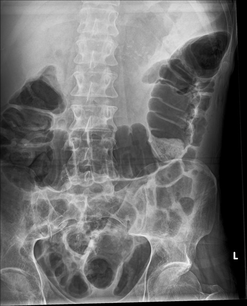
Speckled calcification in the left upper quadrant.
Mass effect on the transverse colon which is displaced inferiorly.
Dilated gas-filled transverse colon, without obstruction, suggests a localized ileus.
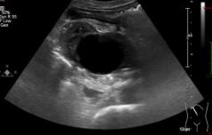
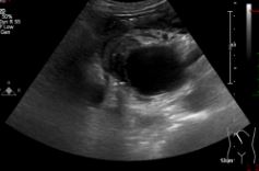
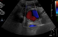
At the region of the head of the pancreas, there is a well-defined, rounded area measuring 4 cm which fills with vascularity on the application of color doppler. This is surrounded by inflammatory soft tissue. Unable to determine the exact nature/source on ultrasound but considering the patient's history of pancreatitis, this may represent pseudoaneurysm.
The liver appears echogenic with a coarse echotexture suggestive of intrinsic liver disease. No focal liver lesion. Antegrade portal venous flow.
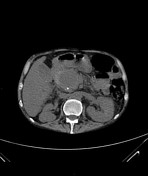

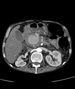

Extensive pancreatic calcification. The pancreas is very atrophic with marked dilatation of the pancreatic duct and marked side branch dilatation.
Large cystic structure in the head of pancreas which is of central low density attenuation with some circumferential high attenuation on the pre-contrast scan, with layering post contrast. This arises from the superior gastroduodenal artery.
No active bleeding.
Moderate intra and extrahepatic duct dilatation with the CBD measuring up to 9 mm. The CBD is compressed by this pseudoaneurysm. Deviation of the duodenum/pylorus and there is some inflammatory stranding.
No focal liver lesions. The spleen, kidneys and adrenal glands are unremarkable.
No free fluid in the abdomen or pelvis.
Case Discussion
Pseudoaneurysms are a recognized complication of severe pancreatitis. This patient was transferred to a tertiary facility and underwent successful coil embolization.


 Unable to process the form. Check for errors and try again.