
Presentation
Suspicion of giant cell arteritis and recent new headaches. No anti-inflammatory treatment. Normal ophthalmological exam.
Patient Data


Axial T1C+ FS scan of the extracranial arteries demonstrates mural thickening and bilateral mural contrast enhancement of the superficial temporal arteries.
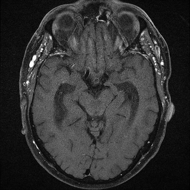
Mild chronic frontal/ethmoidal sinusitis is shown.
Normal presentation of the thoracic vessels.
Case Discussion
Giant cell arteritis (GCA) with typical involvement of the superficial temporal artery is the commonest vasculitis in a Caucasian population. Biopsy is the de facto "gold standard" for diagnosis, however assessment with high resolution contrast enhanced and fat saturated T1- MRI is possible and can also be used for monitoring during/following anti-inflammatory/immunosuppressive therapy. Unlike a biopsy, of course, MRI allows simultaneous assessment of all superficial artery segments.
A symmetrical and simultaneous vascular inflammation of superficial cranial arteries is the typical pattern. MRI imaging signs may vanish after 5-7 days of systemic anti-inflammatory therapy with steroids.
For ranking of mural contrast enhancement, the following four-point scale proposed by Bley et al. can be used:
-, no enhancement; +, slight mural enhancement; ++, prominent mural enhancement; and +++, strong mural enhancement, including perivascular tissue.
NB: for the thickness of the superficial temporal artery wall, the cut-off between unaffected and inflamed arteries is 0.6-0.7 mm.


 Unable to process the form. Check for errors and try again.