
Presentation
Bilateral flank pain and distension.
Patient Data
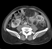

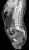
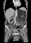
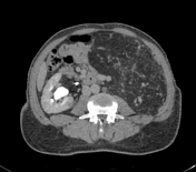

Both kidneys are enlarged in size and show multiple, variable in size, heterogeneously enhancing mass lesions having large fat density and small soft tissue components, the largest one involving the upper half of the left kidney. The largest one exerts mass effects on adjacent organs. These lesions are causing significant loss of the normal parenchyma. No CT evidence of acute haemorrhage is seen. contrast media is normally excreted bilaterally.
The right adrenal gland is unremarkable, however, the left one is not visualised separately.
The liver is mildly enlarged in size with normal position and smooth borders. A small hypodense mass lesion is noted involving segment VI which is showing nodular enhancement on arterial phases and almost complete fill-in on the delayed phase, most likely suggestive of haemangioma.
Note is made of left-sided inguinal hernia and prostatomegaly.
Case Discussion
Current CT findings are most likely suggestive of multiple renal angiomyolipomas on both sides without acute haemorrhage.


 Unable to process the form. Check for errors and try again.